Below the skin in the lower neck is a thick layer of fatty tissue called the scalene fat pad, which helps to protect the deeper muscular and neurovascular structures of the thoracic outlet.
The presence of the scalene fat pad also makes it difficult to feel the structures of the thoracic outlet when pressing with the fingers on the lower neck. Part of the omohyoid muscle, a small muscle that runs across the lower part of the neck, passes through the scalene fat pad.
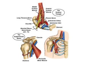
Directly behind the scalene fat pad lies the anterior scalene muscle. This muscle arises from the cervical spine vertebrae and runs vertically to attach to the top of the anterior first rib. Another muscle in this area is the middle scalene muscle. This muscle also arises from the cervical spine vertebrae and runs vertically, attaching to the top of the mid-portion of the first rib. The narrow vertical space formed between the two scalene muscles, with the first rib at the base, is called the scalene triangle. The major nerves and blood vessels serving the upper extremity lie within or adjacent to the scalene triangle as they pass through the upper part of the thoracic outlet over the first rib. The scalene triangle is therefore one of the principal sites for nerve and blood vessel compression that causes TOS.
After passing over the first rib, the brachial plexus nerves, subclavian artery, and subclavian vein all pass together underneath the clavicle. In doing so, they cross underneath a small muscle that runs along the back of the clavicle, called the subclavius muscle. This muscle attaches to the front part of the first rib, anterior and medial to the attachment of the anterior scalene muscle, where it forms a band of dense connective tissue called the costoclavicular ligament. The costoclavicular ligament is immediately lateral to and underneath the sternoclavicular joint. The area between the clavicle and first rib is called the costoclavicular space.
Moving toward the front of the shoulder but before reaching the upper arm, the blood vessels and brachial plexus nerves then pass underneath the pectoralis minor muscle. The pectoralis minor is a relatively small muscle that lies deep to the much larger pectoralis major muscle in the front of the upper chest. It arises from the second, third, and fourth ribs and runs upward to attach to a bony protrusion underneath the clavicle known as the coracoid process. The nerves and blood vessels pass underneath the pectoralis minor tendon through the relatively tight sub-pectoralis space. The sub-pectoralis space is another site for potential nerve or blood vessel compression in TOS.